MAGNETIC MOLECULES & NANOPARTICLES AT SURFACES
People
Valdis Corradini
Alberto Ghirri
Roberto Biagi
Valerio Bellini
Marco Affronte
Introduction.
Low-dimensional magnetism is currently receiving a widespread attention triggered by the technological interest in developing nanomagnets for applications, such as high density data storage, spintronics and quantum information processing. In a realistic device, nanomagnets can be organized on a surface by a rational merging of top-down fabrication (from mm to 50 nm scale) and bottom-up self-assembly (at the nanoscale). The latter, in particular, requires a fine control over the interactions with the surface and between the deposited entities.
Our interest is on deposition and study of molecular magnets and nanoparticles on metal and carbon surfaces. For these systems, the chemical synthesis provides a flexible design of the outer functional shell that, while preserving the pristine properties of the magnetic core, allows to tune their behavior on surfaces.
Experimental methods.
The deposition is carried out by sublimation in ultra-high vacuum (UHV) or simply by drop casting of solutions, while the characterization is performed by means of a UHV variable temperature STM, AFM and spectroscopic techniques (XPS, UPS, LEED, HREELS). Our set-up also includes an electrospray ionization (ESI) source for the (mass filtered) vacuum deposition of thermolabile molecules.
The preservation of the electronic and magnetic properties on surfaces is not a trivial issue. Synchrotron techniques, like X-ray magnetic circular dichroism (XMCD), are regularly employed to carry out these studies. Magnetic nanoparticles are investigated at low temperature by Hall magnetometry, either by scanning Hall probe microscopy (SHPM) or with the direct integration in graphene nanodevices.
 |
|
Fig 1. Schematic of the ESI apparatus. The sample can be positioned at the exit of the mass-filter (pos. S1) or after the prefilter (pos. S2).
|
|
Self assembling of magnetic molecules on gold surface.
|
| |
 |
|
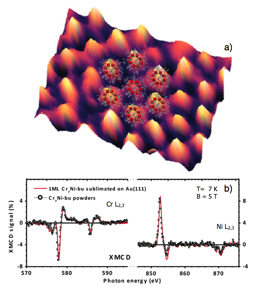
Fig 2. a) STM image showing a hexagonal packed monolayer of Cr7Ni rings on Au(111) with, superimposed to it, the structure calculated by density-functional theory. The sample is obtained by sublimating Cr7Ni powders at 200°C in UHV. b) Comparison between the X-ray magnetic circular dichroism spectra of Cr7Ni taken on pristine powders and monolayer. The excellent agreement of the curves demonstrates the preservation of the magnetic properties at surface.
|
|
Self assembling of magnetic molecules on graphite.
|
| |
 |
|
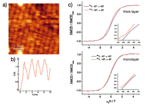
Fig 3. a) STM image of the self-assembled layer formed by Tb double deckers on HOPG as dropcast from a 10-6 M solution in toluene. b) Depth profile measured across four rows of molecules. c) XMCD detected hysteresis of magnetization for the thick layer sample (above) and the monolayer sample (below) on HOPG measured at 7 K. The inset shows the enlargement of the central part of both hysteresis loops.
|
 |
|
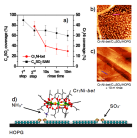
Fig 4. Two steps stabilization of functionalized Cr7Ni-bet rings on HOPG by means of a buffer layer of C16SO3. a) Surface coverage derived by the XPS spectra. AFM images showing b) a homogeneous distribution of molecules on the surface and c) isolated molecules after the rinse with solvent. d) Cartoon illustrating the stabilization mechanism through the formation of a covalent bond between the functional groups of Cr7Ni-bet and C16SO3.
|
 |
|
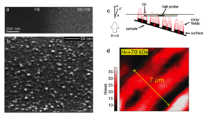
Fig 5. a) SEM images showing the deposition of CsNiCr coordination nanoparticles on a Si substrate functionalized with a suitable ligand. The nanomagnets do not graft on the regions were the ligand has been irradiated by a low dose FIB lithography. b) High magnification image showing the grafted nanoparticles. c) Schematic of the scanning Hall probe microscope. Magnetic stray field are measured by the Hall sensor, which is positioned and scanned on the surface thanks to the tunneling current feedback. d) SHPM image showing the stray field generated by a patterned monolayer stripe of CsNiCr nanoparticles.
|
MAGNETIC X-RAY SPECTROSCOPIES
Experimental
The XAS and XMCD experiments are carried out at the ID8 beamline of the European Synchrotron Radiation Facility (ESRF) in Grenoble (France). The lowest sample temperature reached was about 10 K and the base pressure of the experimental chamber 1.0 x 10⁻¹⁰ mbar. The photon source was an Apple II ondulator that delivers a high flux of polarized light. We paid much attention to avoid any sample degradation induced by radiation exposure, working with very low flux (below 1012 photons/sec) and by strictly monitoring XAS spectra throughout all the experiments for detecting even the smallest traces of sample damaging.
Magnetic dichroism on molecular antiferromagnetic rings.
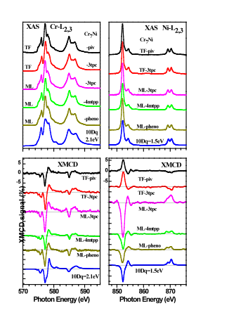
We deeply investigated the properties of submonolayer distributions of isolated molecular Cr7Ni rings deposited on Au(111) by liquid phase (see Fig. 1). X-ray absorption spectra measured at the Cr and Ni L2,3 edges show that the grafting of the Cr7Ni rings onto the gold surface does not affect the oxidation state and the local symmetry of the Cr and Ni sites (see Fig. 2). The circular dichroism shows a change in sign of the Ni magnetic moment. This is due to a reduction in the exchange coupling constants that, however, preserves the structure of the low-energy levels of the grafted rings (see Fig. 4), as corroborated by spin-Hamiltonian simulations and comparison with measurements on bulk sample (see Fig. 3). Density-functional theory calculations show that the Ni-Cr bond gets weaker with slight ring distortion suggesting possible explanation for the observed magnetic behavior. These results show that complex magnetic molecules can be grafted onto surfaces, and that changes in their magnetic behavior must be examined in individual cases.
XMCD measurements at the Cr and Ni L2,3 edges were performed in total electron yield mode using circularly polarized light with about 100% polarization rate and with external magnetic field μ0H up to 5 T applied perpendicularly to the sample surface and parallel to the incident photon beam. The dichroic spectrum is the difference between the XAS spectra taken with the helicity of the incident photon (P) antiparallel (σ↑↓) and parallel (σ↑↑) to the sample magnetization (M). In order to minimize the effects of field inhomogeneity, we first fixed the field and switched the polarization parallel and anti-parallel, then we made the same with the field in opposite direction. The σ↑↑ (σ↑↓) absorption spectra are the mean value of the spectra collected with the helicity parallel (antiparallel) to the sample magnetization.
 |
|
Fig 6. c) STM image (50x25 nm2) and b) 3D view (13x13 nm2) of a single Cr7Ni-3tpc cluster grafted on Au(111) surface. a) Structure of the Cr7Ni-3tpc ring viewed perpendicularly to the Cr7Ni plane.
|
 |
|
Fig 7. Cr (Left panel) and Ni (right panel) L2,3 XAS and XMCD spectra for the Cr7Ni-piv TF, Cr7Ni-3tpc TF and sML, Cr7Ni-4mtpp and Cr7Ni-phenoxy sMLs at 10 K and 5 T, compared with the ligand field multiplet calculations: 10Dq =2.1 eV for Cr3+ and 10Dq=1.5 eV for Ni2+.
|
 |
|
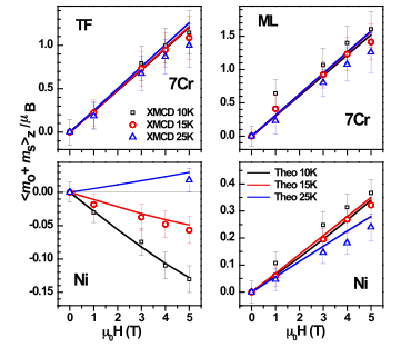
Fig 8. Cr7Ni-3tpc TF (left panel) and sML (right panel). Cr and Ni total magnetic moments, experimentally derived by the sum rules, vs applied magnetic field at 10, 15, and 25 K compared with the results of the spin-Hamiltonian calculations.
|
 |
|
Fig 9. Energy barrier felt by a Cr7Ni-thiobu as a function of the displacement across an Au(111) substrate; the displacement line has been chosen to span different adsorption/symmetry sites for the Au adatom. b) Interaction energy / molecule in the case of a square or hexagonal arrangement of the Cr7Ni-bu molecules as a function of the molecule-molecule distance. c) High resolution STM image, with, superimposed to it, the DFT predicted structure.
|
Magnetic molecules on surfaces: a density-functional approach
To be technologically appealing, molecular magnets must be organized and addressed on solid surfaces or wired to metal electrodes in a controlled way. The first (general) condition in order to be able to adsorb molecules on a surface is obviously that the molecule should endure the deposition procedure. This is rather critical point and the magnetic moment, or even the structural integrity, of very few molecules has been demonstrated so far to survive during deposition in ultra-high vacuum (UHV) on a surface/electrode. Another important point, is how the molecule assembly on the surface, and if, and at which amount, their magnetic properties, are influenced with the interaction with the surface. We simulate by means of density-functional methods the interaction between magnetic molecules and/or their anchoring groups with graphene or metallic surfaces. Information as the adsorption and molecule-molecule interaction energies could be extracted, assisting the experiments (see Figure 8, for and example). In order to treat vdW dispersive forces at some level, semi-empirical corrections to the exchange-correlation potentials, of the Grimme type, have been considered. Real space computer codes as Quickstep, implemented in the CP2K package, and pseudopotentials/PAW codes, e.g. VASP, are used.
Publications
Self-Assembled Monolayer of Cr7Ni Molecular Nanomagnets by Sublimation.
A. Ghirri, V. Corradini, V. Bellini, R. Biagi, U. del Pennino, V. De Renzi, J. C. Cezar, C. A. Muryn, G. Timco, R. E. P. Winpenny, and M. Affronte
ACS Nano 5, 7090 (2011).
Surface Supramolecular Organization of a Terbium(III) Double-Decker Complex on Graphite and its Single Molecule Magnet Behavior.
M. Gonidec, R. Biagi, V. Corradini, M. Moro, V. De Renzi, U. del Pennino, D. Summa, L. Muccioli, C. Zannoni, D. B. Amabilino, and J. Veciana
J. Am. Chem. Soc. 133, 6603 (2011).
Oxo-centered carboxylate-bridged trinuclear complexes deposited on Au(111) by a mass-selective electrospray.
V. Corradini, C. Cervetti A. Ghirri, R. Biagi, U. del Pennino, G. A. Timco, R. E. P. Winpenny, and M. Affronte
New J. Chem. 35, 1683 (2011).
Surface-Enhanced Raman Signal for Terbium Single-Molecule Magnets Grafted on Graphene.
M. Lopes, A. Candini, M. Urdampilleta, A. Reserbat-Plantey, V. Bellini, S. Klyatskaya, L. Marty, M. Ruben, M. Affronte, W. Wernsdorfer, and N. Bendiab
ACS Nano 4, 7531 (2010).
X-ray absorption and magnetic circular dichroism investigation of bis(phthalocyaninato)terbium single-molecule magnets deposited on graphite.
R. Biagi, J. Fernandez-Rodriguez, M. Gonidec, S. Mirone, V. Corradini, F. Moro, V. De Renzi, U. del Pennino, J. C. Cezar, D. B. Amabilino, and J. Veciana
Phys. Rev. B 82, 224406 (2010).
Addressing the magnetic properties of sub-monolayers of single-molecule magnets by X-ray magnetic circular dichroism.
F. Moro, V. Corradini, M. Evangelisti, R. Biagi, V. De Renzi, U. del Pennino, J. C. Cezar, R. Inglis, C. J. Milios, and E. K. Brechin
Nanoscale 2, 2698 (2010).
Deposition of Functionalized Cr7Ni Molecular Rings on Graphite from the Liquid Phase.
A. Ghirri, V. Corradini, C. Cervetti, A. Candini, U. del Pennino, G. Timco, R. J. Pritchard, C. A. Muryn, R. E. P. Winpenny, and M. Affronte
Adv. Funct. Mat. 20, 1552 (2010)
Grafting molecular Cr7Ni rings on a gold surface.
V. Corradini, A. Ghirri, U. del Pennino, R. Biagi, V. A. Milway, G. Timco, F. Tuna, R. E. P. Winpenny, and M. Affronte
Dalton Trans. 39, 4928 (2010).
Successful grafting of isolated molecular Cr7Ni rings on Au(111) surface.
V. Corradini, F. Moro, R. Biagi, V. De Renzi, U. del Pennino, V. Bellini, S. Carretta, P. Santini, V. A. Milway, G. Timco, R. E. P. Winpenny, and M. Affronte
Phys. Rev. B 79, 144419 (2009).
Grafting derivatives of Mn6 single-molecule magnets with high anisotropy energy barrier on Au(111) surface.
F. Moro, V. Corradini, M. Evangelisti, V. De Renzi, R. Biagi, U. del Pennino, C. J. Milios, L. F. Jones, and E. K. Brechin
J. Phys. Chem. B 112, 9729 (2008).
Electronic structure of a Mn6 (S=4) single molecule magnet grafted on Au(111).
U. del Pennino, V. Corradini, R. Biagi, V. De Renzi, F. Moro, D. W. Boukhvalov, G. Panaccione, M. Hochstrasser, C. Carbone, C. J. Milios, and E. K. Brechin
Phys. Rev. B 77, 085419 (2008).
Magnetic Imaging of Cyanide-Bridged Co-ordination Nanoparticles Grafted on FIB-Patterned Si Substrates.
A. Ghirri, A. Candini, M. Evangelisti, G. C. Gazzadi, F. Volatron, B. Fleury, L. Catala, C. David, T. Mallah, and M. Affronte
Small 4, 2240 (2008).
X-ray magnetic circular dichroism investigation of spin and orbital moments in Cr8 and Cr7Ni antiferromagnetic rings
V. Corradini, F. Moro, R. Biagi, U. del Pennino, V. De Renzi, S. Carretta, P. Santini, M. Affronte, J. C. Cezar, G. Timco, and R. E. P. Winpenny
Phys. Rev. B 77, 014402 (2008).